Right coxal bone lateral view – The lateral view of the right coxal bone presents an intriguing canvas upon which we embark on an anatomical journey. From its intricate structure to its pivotal role in human movement, the right coxal bone holds a wealth of knowledge that we unravel in this comprehensive guide.
Prepare to delve into the complexities of the coxal bone, deciphering its bony landmarks, muscular attachments, and potential pathologies. Our exploration unfolds with precision, clarity, and a touch of intrigue, ensuring an immersive learning experience.
Anatomy of the Right Coxal Bone (Lateral View)

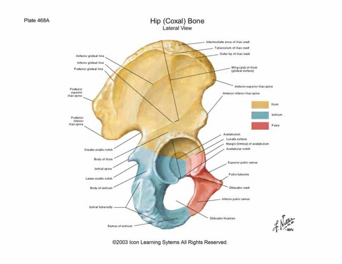
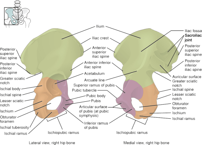
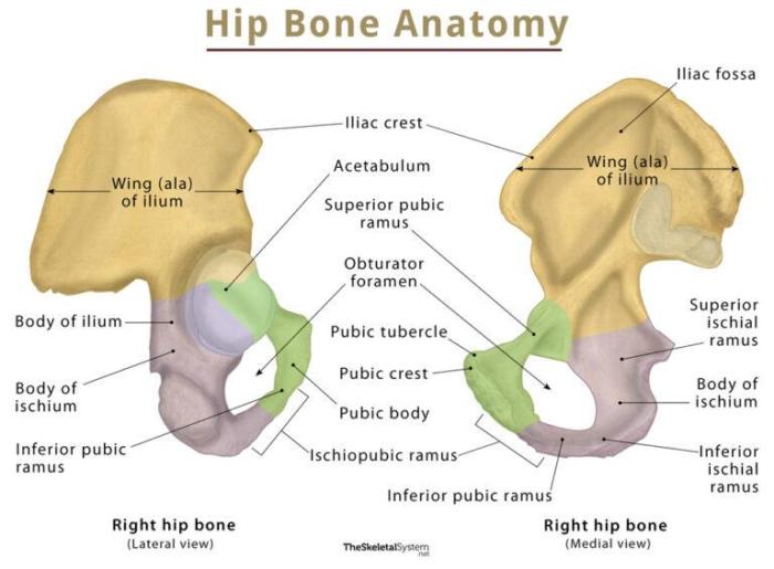
The right coxal bone, also known as the hip bone, is a large, flat bone that forms the lateral wall of the pelvis. It is composed of three fused bones: the ilium, ischium, and pubis. The ilium is the largest of the three bones and forms the upper and posterior portion of the coxal bone.
The ischium is located below the ilium and forms the lower and posterior portion of the bone. The pubis is located anterior to the ischium and forms the anterior portion of the bone.
The coxal bone plays an important role in the human skeletal system. It provides support for the abdominal and pelvic organs, and it helps to transmit weight from the upper body to the lower body. The coxal bone also provides attachment points for muscles that move the hip and leg.
Structure of the Right Coxal Bone (Lateral View), Right coxal bone lateral view
The lateral view of the right coxal bone shows several important anatomical features. The ilium has a large, fan-shaped body that is divided into two parts by a prominent ridge called the iliac crest. The anterior portion of the body is called the iliac fossa, while the posterior portion is called the gluteal fossa.
The iliac crest provides attachment points for muscles that move the hip and trunk. The ischium has a body that is located below the ilium and a ramus that extends anteriorly. The body of the ischium provides attachment points for muscles that move the thigh.
The right coxal bone lateral view provides a detailed look at the structure and landmarks of this important bone. For a step-by-step guide on setting up your printer, refer to lab 10-1 installing a printer . Returning to the right coxal bone lateral view, we can observe the greater sciatic notch and the acetabulum, which are crucial for hip joint function.
The ramus of the ischium forms the inferior border of the obturator foramen, which is a large opening in the coxal bone that allows blood vessels and nerves to pass through.
The pubis has a body that is located anterior to the ischium and a ramus that extends posteriorly. The body of the pubis provides attachment points for muscles that move the thigh. The ramus of the pubis forms the superior border of the obturator foramen.
The pubic symphysis is a joint that connects the two pubic bones at the midline of the body.
Bony Landmarks of the Right Coxal Bone (Lateral View)

The lateral view of the right coxal bone reveals several prominent bony landmarks that serve as crucial references in surgical procedures and diagnostic imaging. These landmarks provide anatomical guidance for surgical interventions and facilitate accurate interpretation of imaging studies.
Major Bony Landmarks
The major bony landmarks visible in a lateral view of the right coxal bone include:
- Ilium:The largest and superior portion of the coxal bone, consisting of the body, ala, and crest.
- Ischium:Located inferior to the ilium, comprising the body, ramus, and tuberosity.
- Pubis:Situated anteriorly to the ischium, formed by the body and superior and inferior rami.
- Acetabulum:A cup-shaped cavity formed by the ilium, ischium, and pubis, which accommodates the head of the femur.
- Greater Trochanter:A large, rounded projection located on the lateral aspect of the proximal femur.
- Lesser Trochanter:A smaller, triangular projection situated inferior to the greater trochanter on the posterior aspect of the femur.
Clinical Significance
The bony landmarks of the right coxal bone are clinically significant in various ways:
- Surgical Procedures:The landmarks serve as anatomical references during surgical interventions, such as hip replacements, pelvic osteotomies, and fracture repairs, guiding the surgeon’s approach and ensuring precise placement of implants.
- Diagnostic Imaging:In radiographs, CT scans, and MRIs, the landmarks assist in accurate interpretation of anatomical structures and identification of pathological conditions, such as fractures, dislocations, and degenerative changes.
| Landmark | Description |
|---|---|
| Ilium | Body, ala, and crest |
| Ischium | Body, ramus, and tuberosity |
| Pubis | Body, superior and inferior rami |
| Acetabulum | Cup-shaped cavity |
| Greater Trochanter | Large, rounded projection |
| Lesser Trochanter | Smaller, triangular projection |
Muscles and Ligaments Associated with the Right Coxal Bone (Lateral View)

The right coxal bone is a complex structure that serves as the foundation for the hip joint. It provides attachment points for numerous muscles and ligaments that contribute to movement, stability, and support of the hip.
Muscles
- Gluteus maximus:The largest muscle of the buttocks, it attaches to the posterior surface of the ilium and extends to the femur. It is responsible for hip extension and external rotation.
- Gluteus medius:Located beneath the gluteus maximus, it attaches to the ilium and greater trochanter of the femur. It abducts the hip and aids in maintaining pelvic stability.
- Gluteus minimus:The smallest of the gluteal muscles, it also attaches to the ilium and greater trochanter. It assists in hip abduction and rotation.
- Tensor fasciae latae:Originating from the ilium and inserting into the iliotibial band, this muscle tenses the fascia lata and contributes to hip abduction and external rotation.
Ligaments
- Iliofemoral ligament:A strong ligament that connects the ilium to the lesser trochanter of the femur. It resists hip extension and internal rotation.
- Ischiofemoral ligament:Extending from the ischium to the greater trochanter, it limits hip extension and external rotation.
- Pubofemoral ligament:Originating from the pubis and attaching to the lesser trochanter, it restricts hip abduction and external rotation.
These muscles and ligaments work together to ensure the stability and mobility of the hip joint, enabling a wide range of movements, including walking, running, and jumping.
Pathologies and Clinical Considerations of the Right Coxal Bone (Lateral View): Right Coxal Bone Lateral View

The right coxal bone is susceptible to various pathologies that can impact its structure and function. These include fractures, dislocations, and arthritis, each presenting with distinct symptoms, diagnosis, and treatment approaches.
Fractures
Fractures of the right coxal bone can occur due to high-impact trauma, such as falls or motor vehicle accidents. Common fracture sites include the acetabulum, ilium, and pubis. Symptoms typically include pain, swelling, bruising, and difficulty walking or bearing weight.
Diagnosis involves physical examination, X-rays, and in some cases, CT scans or MRIs. Treatment options depend on the severity and location of the fracture. Conservative treatment may involve immobilization, pain medication, and physical therapy, while surgical intervention may be necessary for displaced or complex fractures.
Dislocations
Dislocations of the right coxal bone occur when the head of the femur is forced out of the acetabulum. This can happen due to trauma or underlying conditions that weaken the joint. Symptoms include severe pain, deformity, and inability to move the hip.
Diagnosis is made through physical examination and X-rays. Treatment typically involves closed reduction, where the dislocated joint is manipulated back into place under anesthesia. Surgery may be required in cases of complex dislocations or recurrent instability.
Arthritis
Arthritis, particularly osteoarthritis, can affect the right coxal bone, leading to the degeneration of cartilage in the hip joint. Symptoms include pain, stiffness, swelling, and decreased range of motion. Diagnosis involves physical examination, X-rays, and in some cases, MRI scans.
Treatment options aim to manage pain and improve mobility. Conservative measures include pain medication, physical therapy, and lifestyle modifications. In advanced cases, surgical intervention such as hip replacement may be necessary.
Imaging Techniques
Various imaging techniques are used to evaluate the right coxal bone, including:
- X-rays: Provide clear visualization of bones and fractures.
- CT scans: Offer detailed cross-sectional images, helpful for complex fractures or dislocations.
- MRIs: Generate detailed images of soft tissues, ligaments, and cartilage, aiding in the diagnosis of arthritis and other conditions.
Popular Questions
What is the primary function of the right coxal bone?
The right coxal bone, also known as the hip bone, plays a crucial role in weight-bearing, locomotion, and providing stability to the pelvic region.
What are some common pathologies that can affect the right coxal bone?
Common pathologies include fractures, dislocations, and arthritis, which can arise due to trauma, degenerative changes, or underlying medical conditions.
How is the right coxal bone commonly evaluated in clinical settings?
Imaging techniques such as X-rays, CT scans, and MRIs are frequently employed to assess the right coxal bone, aiding in the diagnosis and monitoring of various conditions.